When archeologists discover bones lying deep underground, within ruins, or hidden inside a dark cave, these scientists can use methods like radiocarbon dating to age the bones, or methods like stable isotope analysis to discover the diet of the organism to whom the bones belong. In order to use these methods, the scientists extracts collagen from the bone, and it is with that collagen that the scientist can run the aforementioned tests. Unfortunately, there are many cases when scientists go to extract collagen from a bone and it is not there – over the many years the bones laid dormant within the earth, the collagen escaped the confines of the bone (i.e., decayed). When scientists do find collagen, the extraction process causes damage to the bone and negates the preservation of the artifact.
Recent work by Dr. Maria Squire and colleagues identifies non-destructive methods that can be used by scientists to first determine if collagen is present within a bone before any destructive techniques come into play. Dr. Squire used microCT scanning to take high resolution images of the minuscule structures of bone (see Fig. 1 below). She found that bones with high corticol porosity, that is a large number of holes within the bone, are unlikely to contain enough collagen for analyses like radiocarbon dating and stable isotope analysis (see ET50 in Fig. 1). However, she also found a large amount of variation with the microCT results. So, Dr. Squire and colleagues used additional tests – one being mercury porosimetry – to further test the porosity of the bones. These other techniques can detect pores so small, that they do not show up on microCT images. For example, specimen ET60 (see Fig. 1) appears to lack many pores according to the microCT image, and thus one would expect to find a suitable amount of collagen within the bone for additional analyses; however, this bone does actually contain pores, but these pores are extraordinary small! These minuscule pores were detected by mercury porosimetry, and thus, this bone would be deemed unacceptable for further analyses as there is a good chance it does not contain collagen. In fact, ET60 contained no collagen and if someone tried to extract collagen from it, then they would be destroying the artifact for nothing.
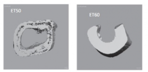
Figure 1. Two microCT scans of bones examined the Tripp et al. paper. Specimen ET50 exhibits a much greater amount of porosity compared to specimen ET60; however, additional analyses revealed small pores throughout ET60 as well.
The destruction of this bone can aid in our on-going quest for knowledge about that organism, but this comes at the cost of the damage done to the bone. The fantastic research by Dr. Squire and her colleagues, however, should limit this destruction as scientists can now use these less invasive techniques to identify suitable bone candidates.
To read more about Dr. Squire’s research, please keep on the lookout for the release of this article “Use of micro-computed tomography imaging and porosity measurements as indicators of collagen preservation in archaeological bone” in the journal Palaeogeography, Palaeoclimatology, Palaeoecology.